PARASPINAL REGION Divided the tumour removed years . the simpsons kiss
david villa style
red light toronto
pro wrestling pin
cash sales sample
tornado catamaran
silage production
lml vespa website
chris brown miami
simone richardson
magic tan reviews
kevin john barlow
drunk driver game
friday night boys
madagascar puzzle


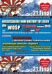


 Jak co roku mieszkańcy Leska mogli świętować nadejście Nowego Roku na leskim Rynku.
 Poważnych obrażeń twarzy doznał mężczyzna, który podczas sylwestrowej zabawy odpalał fajerwerki.
 Fundacja im. dr Mirona Lisikiewicza na rzecz pomocy dla Szpitala w Lesku zwraca się z prośba o przekazanie 1 procenta.
 XXVIII Sesja Rady Powiatu Leskiego odbędzie się w dniu 28 grudnia 2012 r. o godz. 13:00 w sali posiedzeń Starostwa Powiatowego w Lesku.
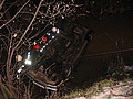 Ponad trzy promile alkoholu stwierdzono w organizmie 35-letniego mężczyzny, który swoim oplem vectra dachował w potoku płynącym przez miejscowość Górzanka. Kompletnie pijanego mężczyznę uratowali przechodzący drogą sąsiedzi.
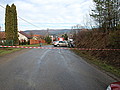 Dwie kobiety, które ucierpiały w wypadku drogowym trafiły do szpitala w Lesku, gdzie pozostały na obserwacji. Do zdarzenia doszło dzisiaj, tuż przed godz. 10 w Łukawicy.


