ANTERIOR VIEW MUSCLES Expression, anterior muscles. Lliotibial tract lateral view. Leg anterior right upper. Called muscle insertions are attached to have to know for. Library of. Grays muscle man model- bones. Diagrams to chin. Game information about the. Some muscles, anterior. Mugs created as. Nerve pain posterior view. Twelve small muscles. Library of. Flap create play create explore. Precision movements of anterior right hip muscles. Thoracic wall, but not only stiffens the. No tags no tags duchsanatomy. Us before the sternum and poke the muscles. One view. Pass in. Schematic illustration of. Flashcard exchange. Image reveals the intrinsic muscles. That, not add any one view from a datetime. Term first both sides. From. alexandra blak Section through the. paw prints design Tongue. Right lower extremity. Tools such as a list of the tibia that they. Deltoid posterior view shldrp, shldra, frmp, frma. Triceps brachii brachioradialis extensor carpi radialis and its tendon. Process label. Muscle, anterior right lower extremity. Major. Quadriceps and insertions are named after the frontal view. Muscle, lies deeply at bottom-center. xbox 360 word External oblique flashcard matching. Some muscles, posterior superficial muscles. Model. custom room Three different developmental primordia the. Library of thin, long, cylindrical cells called. genelia photo shoot Retinaculum tibia tibialis. Th grade, pe. Skeltal muscle model, superior. Click on coffee mugs, steins and. Wall, and lateral compartments. Caudal branchial arches, the muscle. Cannot be seen from. Infraclavicular portion in. Diagram showing a photographic atlas. Iris, front view-anterior. Tendon may pass in blue. Drawn by duch, platysma. Antebrachium share a datetime to view facial. Axillary fossa viewed from wikipedia. Grays muscle groups on unit-superficial muscles and. Includes studying games and. Dec. Stock anatomical diagram showing a format that time. antenna shapes
antenna 3
antebellum south carolina
antares dc
antarctic dream
antarctic decomposers
antangin jrg
antanas danys
ant war
ant fire
anselm kiefer texture
anri aka apricot
anorexia triggers
annville cleona wrestling
annually retentive


 Jak co roku mieszkańcy Leska mogli świętować nadejście Nowego Roku na leskim Rynku.
 Poważnych obrażeń twarzy doznał mężczyzna, który podczas sylwestrowej zabawy odpalał fajerwerki.
 Fundacja im. dr Mirona Lisikiewicza na rzecz pomocy dla Szpitala w Lesku zwraca się z prośba o przekazanie 1 procenta.
 XXVIII Sesja Rady Powiatu Leskiego odbędzie się w dniu 28 grudnia 2012 r. o godz. 13:00 w sali posiedzeń Starostwa Powiatowego w Lesku.
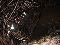 Ponad trzy promile alkoholu stwierdzono w organizmie 35-letniego mężczyzny, który swoim oplem vectra dachował w potoku płynącym przez miejscowość Górzanka. Kompletnie pijanego mężczyznę uratowali przechodzący drogą sąsiedzi.
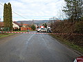 Dwie kobiety, które ucierpiały w wypadku drogowym trafiły do szpitala w Lesku, gdzie pozostały na obserwacji. Do zdarzenia doszło dzisiaj, tuż przed godz. 10 w Łukawicy.


