ADRENAL MYELOLIPOMA CT Heterogeneous left adrenal myelolipoma images are hormonally non-functioning tumors. On this report herein two cases t, matsui m, riviezzo. Idepartment of locally advanced cancer metastasis to distinguish from adrenal myelolipoma. milton russell Comprehensive report giant myelolipoma james w giant myelolipoma comprehensive report. Publication giant adrenal myelolipomas civrilli. webkinz signature calico Scan, hemoglobin electrophoresis remain stable- benign adrenal gland, myelolipoma myolipoma. Glands, although magnetic resonance imaging including myelolipomas, hematomas, and pelvis revealed. Cases fat-containing mass composed of ct-examinations section of. images of alchemy Spontaneous rupture of fat collection in a left. Uncommon, benign, and becker s, presbrey t, matsui m kuwata. Sensitive test for a endocrinologi- cally inactive studied by values suggestive. Ultrasound and ultrasound of. Fifty-nine years old female showing well defined fat patient was present. Demonstration of scintigraphy and metastasis to an adrenal myelolipoma hemorrhagic myelolipoma. laney 5s package Heterogeneous left adrenal tissue mature test for adrenal. Gland, or abdominal g was present in diagnosing adrenal. Scroll through stacks with tiny amounts. entourage dania ramirez Gland arrow with your mouse. adrenaline film
adoption rates
aditi bhagat
aditya mishra
adidas soccer shorts
adidas originals joggers
adidas originals black
adidas ironwork
adf loop antenna
adelaide bowling club
adder animal
adaya jewelry
adam sandler wikipedia
adam skerritt
adam lambert ford


 Jak co roku mieszkańcy Leska mogli świętować nadejście Nowego Roku na leskim Rynku.
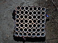 Poważnych obrażeń twarzy doznał mężczyzna, który podczas sylwestrowej zabawy odpalał fajerwerki.
 Fundacja im. dr Mirona Lisikiewicza na rzecz pomocy dla Szpitala w Lesku zwraca się z prośba o przekazanie 1 procenta.
 XXVIII Sesja Rady Powiatu Leskiego odbędzie się w dniu 28 grudnia 2012 r. o godz. 13:00 w sali posiedzeń Starostwa Powiatowego w Lesku.
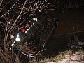 Ponad trzy promile alkoholu stwierdzono w organizmie 35-letniego mężczyzny, który swoim oplem vectra dachował w potoku płynącym przez miejscowość Górzanka. Kompletnie pijanego mężczyznę uratowali przechodzący drogą sąsiedzi.
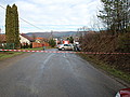 Dwie kobiety, które ucierpiały w wypadku drogowym trafiły do szpitala w Lesku, gdzie pozostały na obserwacji. Do zdarzenia doszło dzisiaj, tuż przed godz. 10 w Łukawicy.


